
(2014) Ultrastructure of spermatozoa of orsolobidae (Haplogynae, Araneae) with implications on the evolution of sperm transfer forms in Dysderoidea. Arthropod structure & Development, 44, 218–227. (2015) The Last Breath: A μCT-based method for investigating the tracheal system in Hexapoda. (2002) Head structures of Priacma serrata leconte (coleptera, archostemata) inferred from X‐ray tomography. Journal of Apicultural Research, 47 (4), 286–291. (2008) X-ray computerised microtomography (MicroCT): a new technique for assessing external and internal morphology of bees. (2017) X-Ray microtomography for ant taxonomy: An exploration and case study with two new Terataner (Hymenoptera, Formicidae, Myrmicinae) species from Madagascar. Garcia, F.H., Fischer, G., Liu, C., Audisio, T.L., Alpert, G.D., Fisher, B.L. (2014) Insect morphology in the age of phylogenomics: Innovative techniques and its future role in systematics. 70781U., San Diego, California, USA.įriedrich, F., Matsumura, Y., Pohl, H., Bai, M., Hörnschemeyer, T. Proceedings of Society of Photo-Optical Instrumentation Engineers (SPIE) 7078, Developments in X-Ray Tomography VI. (2008) Micro-computer tomography and a renaissance of insect morphology. (2016) Revision and Microtomography of the Pheidole knowlesi Group, an Endemic Ant Radiation in Fiji (Hymenoptera, Formicidae, Myrmicinae). (2013) Micro-computed tomography : Introducing new dimensions to taxonomy. įaulwetter, S., Vasileiadou, A., Kouratoras, M., Dailianis, T. (2015) A review of the subfamily Rogadinae (Hymenoptera: Braconidae) from Iran. įarahani, S., Talebi, A.A., van Achterberg, C. (2012) First records of Macrocentrus Curtis, 1833 (Hymenoptera: Braconidae: Macrocentrinae) from Northern Iran. Entomological Magazine, 1, 186–199.įarahani, S., Talebi, A.A. (1833) Characters of some undescribed genera and species indicated in the "Guide to an arrangement of British insects. Journal of Hymenoptera Research, 18, 1–24.Ĭurtis, J. (2009) Structure and functional morphology of the ovipositor of Homolobus truncator (hymenoptera: Ichneumonoidea: Braconidae). (2008) On the head morphology of Tetraphalerus, the phylogeny of Archostemata and the basal branching events in Coleoptera. Zoological Journal of Linnean Society, 158 (3), 533–562. (2010) The larval head of Nevrorthidae and the phylogeny of Neuroptera (Insecta). Journal of Insect Biodiversity and Systematics, 1 (2), 111–123.īeutel, R.G., Friedrich, F. (2016) A Review of the Subfamily Hormiinae (Hymenoptera: Braconidae) from Iran. (2018) New avatars for myriapods: Complete 3D morphology of type specimens transcends conventional species description (Myriapoda, Chilopoda). (2015) A new dimension in documenting new species: high-detail imaging for myriapod taxonomy and first 3D cybertype of a new millipede species (Diplopoda, Julida, Julidae).
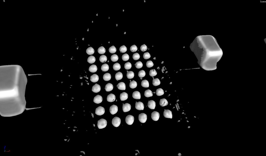
#MICROCT VIDEO ROTATE 3D RECONSTRUCTED IMAGE DOWNLOAD#
The complete datasets containing the raw TIFF MicroCT data, 3D models and 3D rotation videos available for download at Īkkari, N., Enghoff, H. As a result, the use of micro-computed tomography as a non-invasive virtual examination tool was explored. A brief step-by-step is provided on image acquisition, 3D reconstruction and mesh editing to create a virtual model of the species utilized for morphological and morphometric studies. MicroCT scan data of three braconid wasp species from Iran, depicted main identification of skeletal body parts. In this study, MicroCT imaging protocols are explained in detail using three species of braconid wasps: Aleiodes arnoldii Tobias, 1976 (Braconidae: Rogadinae), Hormius moniliatus Nee, 1811 (Braconidae: Hormiinae) and Macrocentrus bicolor Curtis, 1833 (Braconidae: Macrocentrinae). However, this technology is rarely used in entomology compared to in medical and industrial applications. Micro-computed tomography, as a non-destructive imaging technique, has become an emerging and progressive technology in insect science. New technologies for imaging and analyzing in taxonomy, offer opportunities to deposit three-dimensional (3D) data to proposed for rare and valuable type materials in museums and collections. Traditionally, entomologists have used destructive methods especially dissection in order to investigate the important taxonomic characters of specimens. Micro-CT, 3D imaging, Cybertype, Braconidae, taxonomy, parasitoids Abstract Institute for Photon Science and Synchrotron Radiation (IPS), Karlsruhe Institute of Technology (KIT), Hermann-von-Helmholtz-Platz 1, Eggenstein-Leopoldshafen, Germany. Department of Agricultural Entomology, Faculty of Agriculture, Tarbiat Modares University, Tehran, Iran.


 0 kommentar(er)
